Datei:Schematic representation of the hypothalamic nuclei.png
aus Wikipedia, der freien Enzyklopädie

Größe dieser Vorschau: 800 × 342 Pixel. Weitere Auflösungen: 320 × 137 Pixel | 640 × 274 Pixel | 1.276 × 546 Pixel.
Originaldatei (1.276 × 546 Pixel, Dateigröße: 539 KB, MIME-Typ: image/png)
Dateiversionen
Klicke auf einen Zeitpunkt, um diese Version zu laden.
| Version vom | Vorschaubild | Maße | Benutzer | Kommentar | |
|---|---|---|---|---|---|
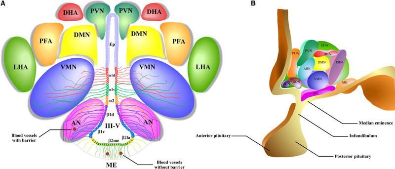
| aktuell | 15:41, 15. Sep. 2018 |  | 1.276 × 546 (539 KB) | wikimediacommons>Was a bee | {{Information |Description={{en|1=A schematic representation of the hypothalamic nuclei and the distribution of tanycytes over the wall of the third ventricle (III-V). (A) Coronal view of the approximate location of the hypothalamic nuclei and tanycytes. Ciliated ependymocytes (ep) line the dorsal wall of the III-V. The α1d-tanycytes (α1d) and α1v-tanycytes (α1v) have long projections that make contact with the neurons of the VMN. α2-tancycytes (α2) have projections to the AN and blood vessel... |
Dateiverwendung
Die folgende Seite verwendet diese Datei:
